You’ll find that bird legs are mainly made up of three key bones: the femur, tibiotarsus, and tarsometatarsus. The femur connects the hip to the knee, while the tibiotarsus is a fusion of the tibia and ankle bones. The tarsometatarsus links the ankle to the toes.
Together, these fused bones provide strength, flexibility, and specialized movement. Understanding these will offer insights into how birds plunge, walk, and adapt to diverse environments.
Key Takeaways
- Bird legs mainly consist of the femur, tibiotarsus, and tarsometatarsus bones.
- The femur connects the hip to the knee and is shorter and robust for bipedal movement.
- The tibiotarsus links the knee to the foot, formed by fused tibia and upper foot bones.
- The tarsometatarsus is a fusion of distal tarsal bones and metatarsals, supporting walking and perching.
- Toe arrangements vary widely, influencing bird movement and grip, with common types like anisodactyl and zygodactyl.
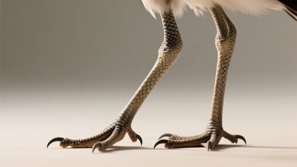
Basic Structure of Bird Legs
The basic structure of bird legs centers on two main bones: the tibiotarsus and the tarsometatarsus. Both result from the fusion of multiple leg and foot bones to support efficient bipedal movement. The tibiotarsus forms by fusing the tibia with upper foot bones, while the tarsometatarsus comprises fused distal bones plus metatarsals II, III, and IV.
You won’t see the bird’s knee externally because it’s concealed close to the body beneath feathers. The tarsus, the segment between the ankle and toes, plays an essential role in connecting the foot to the leg.
Bird legs also display diverse adaptations in toe arrangement and foot structure, tailored to their ecological functions like perching or swimming. This highlights the specialized morphology vital for their varied locomotion.
Femur: The Upper Leg Bone
Bird femurs serve as the critical link between the hip and knee joints, enabling a wide range of motion essential for various movements.
You’ll notice the femur in birds is shorter and more robust than in mammals, an adaptation for efficient bipedal locomotion.
At its proximal end, the femur forms a ball-and-socket joint with the hip, granting birds exceptional mobility.
This unique structure also contributes to their lightweight skeleton, which is crucial for flight.
The femur often fuses with other leg bones, enhancing limb strength and efficiency during walking and running.
Understanding the femur’s specialized design helps you appreciate how birds balance strength, flexibility, and lightweight construction in their upper leg bones.
Tibiotarsus: Fusion of Tibia and Ankle Bones
Following the femur’s role in connecting the hip to the knee, the tibiotarsus extends the leg by linking the knee joint to the foot. The tibiotarsus forms through the fusion of the tibia and proximal tarsal bones, creating a single, elongated structure that improves leg stability during movement.
At its proximal end, the cnemial crest anchors powerful leg muscles essential for running and jumping. Unlike mammals, birds lack separate centralia bones within the tibiotarsus, simplifying their hind limb anatomy.
This fusion plays a significant role in avian bipedalism, allowing for efficient locomotion and adaptability across diverse environments.
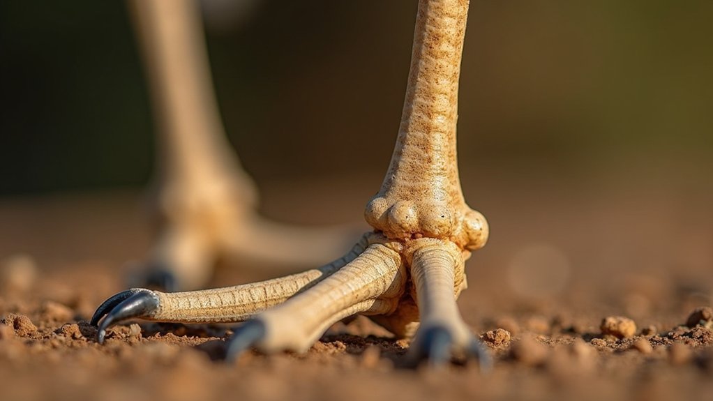
Tarsometatarsus: The Lower Leg and Foot Fusion
You’ll find that the tarsometatarsus forms through the fusion of distal tarsal bones and metatarsals II, III, and IV, creating a single, robust structure.
This fusion really boosts the bird’s foot strength and flexibility, which in turn directly affects how it moves and perches.
When you look at variations in the shape and length of this bone, you can see evolutionary adaptations that have been fine-tuned for different ecological needs.
It’s pretty fascinating how these changes help birds thrive in their specific environments.
Bone Fusion Explained
Although the fusion of bones in the avian lower leg may seem complex, understanding the tarsometatarsus is essential for grasping how birds achieve their distinctive locomotion. This bone results from the fusion of distal tarsals and metatarsals II, III, and IV, creating a single elongated structure that supports the foot.
The first metatarsal remains separate, anchoring the first toe for balance. Unlike mammals, birds lack individual ankle and foot bones, optimizing their walking and running. This fusion modifies leg mechanics, enhancing bipedal efficiency.
| Bone Component | Description |
|---|---|
| Distal Tarsals | Fused with metatarsals II-IV |
| Metatarsals II-IV | Form the main shaft of tarsometatarsus |
| Metatarsal I | Remains separate, supports first toe |
| Tarsometatarsus | Single elongated fused bone |
| Absence of Centralia | Unique adaptation in bird legs |
Structure and Function
Because the tarsometatarsus fuses distal tarsal bones with metatarsals II, III, and IV, it forms a robust lower leg and foot structure that efficiently distributes weight and augments mobility.
You’ll find that this fusion improves the bird’s ability to walk, run, and perch by providing a stable yet flexible support system.
The tarsometatarsus connects proximally to the tibiotarsus, itself a fusion of the tibia and upper foot bones, which further strengthens the leg’s framework.
Significantly, the first metatarsal remains separate, anchoring the hallux and allowing diverse toe arrangements critical for various functions.
Evolutionary Adaptations
When examining the evolutionary adaptations of bird legs, you’ll notice that the fusion of distal tarsal bones with metatarsals II, III, and IV into the tarsometatarsus plays a significant role. This fusion forms a single, robust bone that improves strength and stability in the lower leg, essential for efficient bipedal locomotion.
By reducing the number of bones, the tarsometatarsus supports powerful movements such as walking, running, and perching.
Alongside the tibiotarsus, formed by fusing the tibia with upper foot bones, this adaptation optimizes force transmission during take-offs and landings.
The evolution of these fused structures in bird feet reflects adaptations for diverse environments and specialized locomotion, highlighting their importance in avian mobility and flight-related functions.
Understanding the Patella and Cnemial Crest
If you’ve ever examined bird anatomy closely, you’ll notice that the patella, or kneecap, sits just above the cnemial crest on the tibia. However, some bird species don’t have a patella at all. The cnemial crest acts as a significant muscle attachment site, enhancing leg movement efficiency.
This anatomical relationship is essential for understanding bird locomotion mechanics.
| Feature | Function |
|---|---|
| Patella | Protects the knee joint; improves advantage |
| Cnemial Crest | Muscle attachment; aids in leg extension |
| Tibia | Main bone of the lower leg |
| Absence of Patella | Observed in some species; affects movement |
Grasping this connection helps you appreciate how birds optimize their legs for varied functions like perching and running.
The Role and Reduction of the Fibula
Although the fibula in birds is significantly reduced compared to other vertebrates, it plays an essential supportive role by closely adhering to the tibia rather than bearing weight.
In most bird species, the fibula doesn’t extend the full length of the tibia, reflecting its diminished structural importance. This reduction contributes to a lightweight skeletal design, which is vital for flight efficiency.
Importantly, penguins retain a full-length fibula, adapted for swimming rather than terrestrial locomotion.
The fibula’s position beneath the well-developed cnemial crest highlights its role as an attachment site for powerful locomotor muscles. By providing this support without significant weight-bearing, the fibula balances structural integrity with the demands of avian mobility and flight adaptations.
Understanding this reduction clarifies how bird leg anatomy varies from that of mammals.
Distinguishing the Knee From the Ankle
Understanding the fibula’s role helps clarify the complex arrangement of bird leg joints, which often leads to confusion between the knee and ankle.
In birds, what looks like a knee is actually the ankle or heel joint, typically concealed beneath feathers. The true knee lies above the cnemial crest and remains hidden from view in most species. This knee joint connects the femur to the tibiotarsus, unlike the ankle, which links the tibiotarsus to the tarsometatarsus.
Since birds walk on their toes, the tarsometatarsus acts as a critical bone between the ankle and toes.
Recognizing the knee’s position close to the body and distinguishing it from the visible ankle is essential for understanding avian locomotion and leg anatomy accurately.
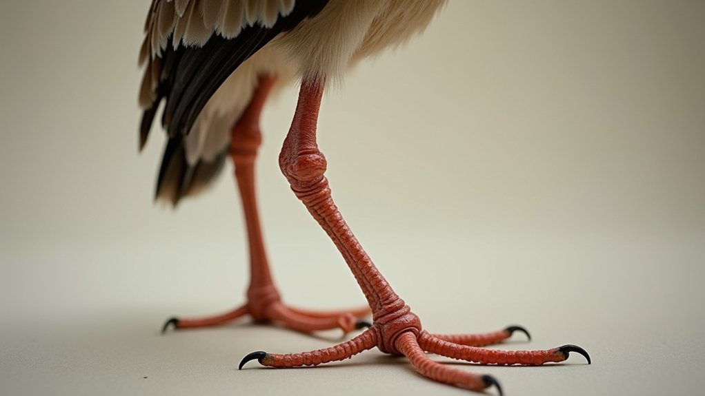
Toe Arrangements and Their Functions
You’ll notice that birds have distinct toe arrangements like anisodactyl, zygodactyl, and didactyl.
Each of these is tailored to specific behaviors—whether it’s perching, climbing, or running. These configurations really optimize their grip and mobility, which helps them survive in all sorts of environments.
Common Toe Arrangements
Toe arrangements in birds play an essential role in their locomotion and interaction with the environment. They directly influence their ability to perch, climb, run, or swim.
The most common arrangement is anisodactyl, where three toes face forward and one toe, the hallux, points backward. This setup maximizes stability for perching.
Zygodactyl feet, with two toes forward and two backward, appear in woodpeckers and parrots. This helps them grip vertical surfaces better.
Some woodpeckers have tridactyl feet, featuring three forward-facing toes suited to their habitat.
Ostriches possess didactyl feet, with only two toes, optimized for swift running on open terrain.
Moreover, heterodactyl feet, unique to trogons, and syndactyl feet, where two toes are fused as in kingfishers, further diversify bird toe configurations.
These variations reflect their ecological niches.
Functional Adaptations
A bird’s toe arrangement directly influences its interaction with the environment, determining how it perches, climbs, or swims. You’ll notice birds’ feet adapt specifically to their lifestyle: anisodactyl toes stabilize perching; zygodactyl toes grip trunks; syndactyl toes aid swimming. These specialized adaptations guarantee survival and efficiency.
| Toe Arrangement | Function | Emotion Evoked |
|---|---|---|
| Anisodactyl | Stable perching | Calm, grounded |
| Zygodactyl | Strong gripping | Determined, focused |
| Syndactyl | Swimming efficiency | Graceful, fluid |
Understanding these details helps you appreciate how birds’ feet perfectly match their ecological niches. It highlights nature’s precision in evolution.
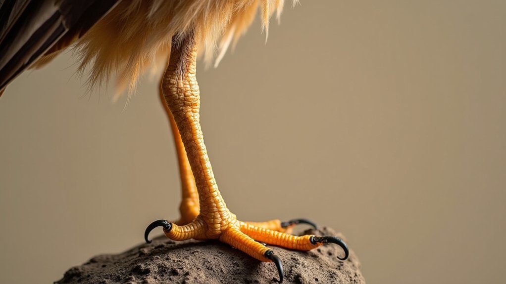
Claws and Their Adaptations
Although bird claws share the common function of covering the distal ends of their toes, their curvature, size, and structure vary considerably to meet species-specific needs.
You’ll notice that larger birds often possess claws with a greater radius of curvature, which improves grip and stability during perching or hunting.
Raptors exemplify this adaptation with sharp, curved talons designed for seizing prey efficiently. Claws also play an essential role in climbing; species like woodpeckers have strong, pointed claws that enable secure attachment to tree bark.
Conversely, aquatic birds such as ducks exhibit less pronounced claws, often with webbing, prioritizing swimming over grasping.
Understanding these variations highlights how claws are specialized tools finely tuned to support diverse avian lifestyles and ecological niches.
Webbing, Lobation, and Swimming Adaptations
Three primary types of foot adaptations—webbing, lobation, and partial webbing—equip aquatic birds to navigate their watery environments efficiently.
Palmated feet, with webbing connecting the anterior digits, allow you to paddle effectively through water.
Totipalmate feet take this further: webbing connects all four toes, providing powerful propulsion seen in gannets and pelicans.
Semipalmate feet feature partial webbing between the front toes, balancing swimming and walking on soft substrates, common in plovers and sandpipers.
Lobate feet, distinguished by skin lobes edging the anterior digits, improve swimming by increasing surface area without full webbing. You can observe this in species like the American coot.
Understanding these structural variations helps you appreciate how webbing and lobation optimize aquatic locomotion across bird species.
Thermal Regulation in Bird Feet
When birds stand on icy surfaces, they use countercurrent exchange systems in their feet to minimize heat loss by transferring warmth from arterial to venous blood. This mechanism allows the legs and feet to conserve core body heat efficiently.
For example, gulls constrict blood vessels in their feet, reducing blood flow and further limiting thermal loss. Conversely, some species can bypass this heat exchange to dissipate heat during hot conditions, aiding in temperature regulation.
The feet of ducks and geese feature specialized adaptations that prevent freezing, enabling them to swim in cold water without frostbite.
Thermal regulation in bird legs and feet is essential for survival, especially in extreme climates. It maintains activity levels and prevents hypothermia by balancing heat retention and dissipation with precision.
Frequently Asked Question
How Do Bird Leg Muscles Differ From Mammal Leg Muscles?
You’ll find bird leg muscles differ from mammal leg muscles by being specialized for rapid, powerful movements essential for flight and take-off.
Birds have more fast-twitch fibers, making their muscles lighter and streamlined for aerodynamic efficiency.
Mammal leg muscles are bulkier, designed for diverse functions like running or climbing, with a mix of fiber types.
Furthermore, bird muscles adapt to fused bones, enhancing precise toe control for perching, unlike mammals.
What Types of Feathers Cover a Bird’s Legs?
In regard to feathers on bird legs, you’ll mainly find contour and down feathers covering them, providing insulation and protection.
Some birds, like raptors, have specialized feathers that aid in gripping prey and add extra defense.
Waterfowl sport waterproof feathers on their legs, helping keep them dry while swimming.
In contrast, ostriches and emus have bare, scaly skin to help with heat dissipation, showing nature’s way of covering all bases.
How Do Bird Legs Heal After Injury?
When a bird’s leg gets injured, its bones heal efficiently thanks to a rich blood supply and rapid cellular activity.
You’ll notice a callus forms around the fracture, stabilizing the area as new bone tissue replaces it.
Birds also use their feathers for insulation and infection protection.
By resting and limiting movement, they aid recovery.
Depending on species and injury severity, healing can take two to six weeks.
Can Bird Leg Anatomy Indicate Their Habitat or Diet?
Just like Sherlock Holmes reads clues to solve mysteries, you can analyze bird legs to understand their habitat and diet.
Long, slender legs suggest wading in shallow waters, while robust, sturdy legs indicate ground feeding and running.
Webbed feet reveal aquatic lifestyles, and zygodactyl toes point to climbing habits.
How Do Bird Leg Bones Grow and Develop Over Time?
You’ll find bird leg bones grow through the fusion of multiple bones, like the tibiotarsus and tarsometatarsus, which reduces weight and improves movement.
Genetic and environmental factors influence this development, allowing adaptation to different habitats.
Hormonal changes regulate ossification, strengthening the bones as the bird matures.
The fibula often reduces, except in penguins, reflecting evolutionary optimization for flight and mobility, with bone size and shape adapting to specific functions.
Conclusion
You might think bird legs are just simple limbs, but they’re complex structures distinctly adapted for various functions, from perching to swimming. Understanding terms like tibiotarsus and tarsometatarsus reveals how evolution fuses bones for strength and efficiency.
These specialized features aren’t just anatomical curiosities; they’re essential for survival. So next time you see a bird, remember its legs are marvels of natural engineering, perfectly designed for its lifestyle and environment.